
Home-Based Hypertension Program Produces ‘Striking’ Results

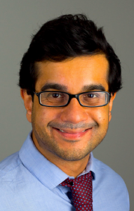
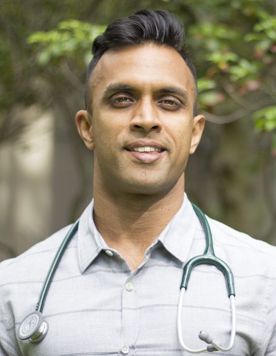
Naomi Fisher
A pilot study conducted by Brigham investigators found that an innovative care-delivery program helped participants reduce their blood pressure in just seven weeks.
Hypertension, or high blood pressure, affects nearly half of U.S. adults, according to the American Heart Association. Despite the serious consequences that can result from hypertension—which puts patients at an increased risk for heart attacks, strokes and other cardiovascular events—elevated blood pressure often remains untreated or undertreated for years.
Seeing opportunities for improvement, a team of innovators and clinicians at the Brigham developed a new care-delivery program for home use that aims to improve hypertension control rates quickly and at a significantly lower cost than traditional, clinic-based blood pressure programs.

The new approach, piloted with 130 participants, helped 81 percent of patients achieve blood pressure control in, on average, less than two months. The results of the pilot study were published recently in Clinical Cardiology. Patients who enrolled had uncontrolled blood pressure (greater than 140/90) and were recruited from Brigham and Women’s Primary Care Associates, Longwood, and the Watkins Cardiovascular Clinic.
“This is a striking result, especially given the very short time frame in which control was reached,” said Naomi Fisher, MD, director of the Brigham’s Hypertension Service and Hypertension Specialty Clinic. “There are a few notable health care systems that have matched or exceeded this control rate, but most clinical practices do not approach this rate of success.”
Incorporating Technology
To overcome some of the challenges that clinical practices face, Fisher and colleagues combined several innovative strategies to create their program. Participants each received a Bluetooth-enabled blood pressure device that automatically transmits blood pressure measurements—taken by patients at home—into electronic medical records.
In addition, patients had access to “patient navigators”—non-physicians trained to use a clinical algorithm developed by hypertension specialists. The program enabled rapid assessment and medication dosage adjustments for patients. Medication adjustments were made every two weeks until home blood pressure was under 135/85.
The next step will be to test the program’s effectiveness with a larger population. With this approach, the team anticipates significant cost effectiveness and cost savings, in addition to the prevention of cardiovascular events and death from treating hypertension more intensively in men and women.
“The time-honored model of treating hypertension via traditional visits to the doctor is neither effective nor sustainable,” said Fisher. “Organizations can and should develop and adopt innovative technologies to create sustainable solutions for hypertension control.”
This work was supported by an institutional Brigham Care Redesign Incubator and Startup Program (BCRISP) award.
 To examine these trends, hospital discharge data was acquired from Massachusetts and Florida between 2007 and 2013 and from California between 2007 and 2011. This information came from the State Inpatient Databases (SIDs) of the Healthcare Cost and Utilization Project, created by the federal Agency for Healthcare Research and Quality. This dataset includes information such as homeless status, billing, demographics and diagnoses.


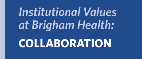
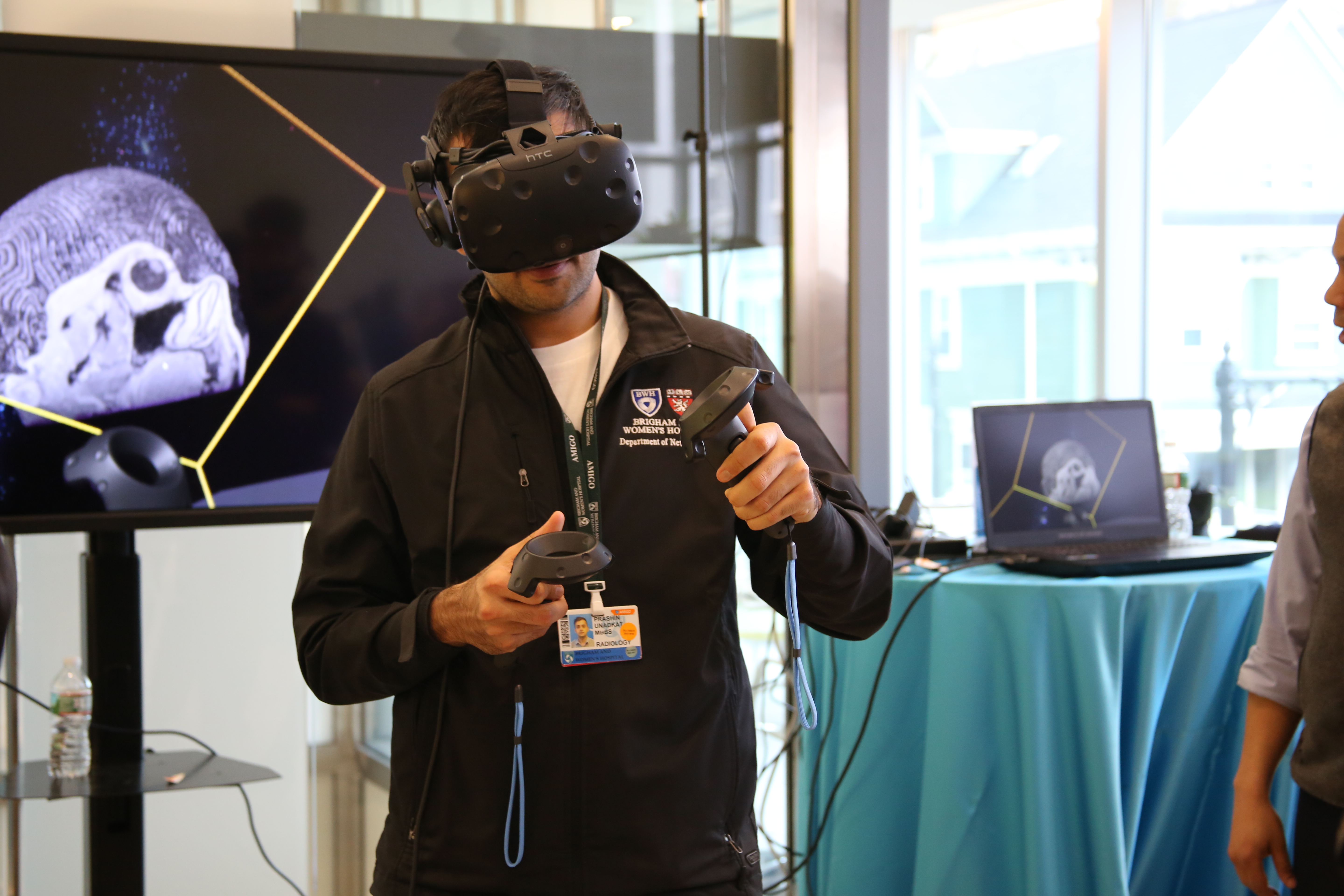
 Brigham Health’s Strategy in Action: Discovery and Innovation


 “When I got that diagnosis, it hit me like a brick. I thought, wow, there’s something else going on,” she said. “Genetically speaking, there’s no explanation for it.”


 “Our staff’s commitment to service excellence, professionalism and team spirit came through loud and clear to the surveyors,” said Milenko Tanasijevic, MD, MBA, vice chair for Clinical Pathology and Quality. “They go above and beyond for every specimen and demonstrate their dedication to our patients every single day, regardless of whether a survey is occurring.”
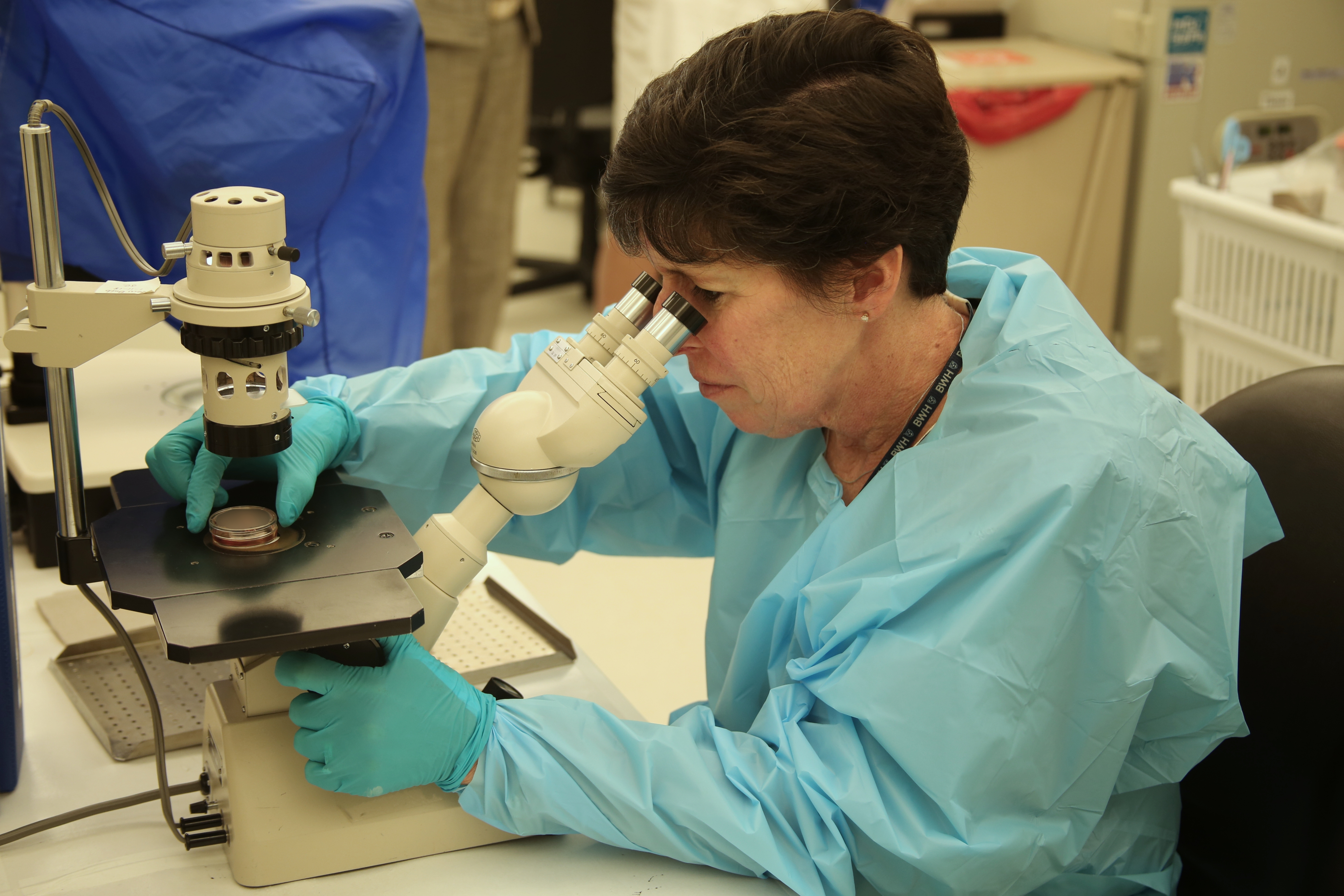


 The following year, Sinha joined a panel to judge the 2015 finalists for the Stepping Strong Innovator Awards competition. That’s where he heard BWH bioengineer Su-Ryon Shin, PhD, present her work with 3-D bioprinting. Sinha sought her out immediately after the presentation, and the two started talking about how they could help one another – and, ultimately, patients who had sustained muscle trauma injuries. Their conversation has continued ever since. In 2017, they received an additional Stepping Strong grant of $100,000 to join forces to continue their work together.


 Mohamed El-Dib, MD, director of Neonatal Critical Care in the Department of Pediatric Newborn Medicine, is the principal investigator at BWH for a multi-institutional clinical trial looking at whether infusing babies with their own umbilical cord blood can indeed reverse tissue damage in the brain.
 Radiology images can reveal critical clues not only about a patient’s current injuries, but also about patterns of violence a person may be experiencing. A recent study by BWH investigators reveals new ways that radiologists can be involved in the care of victims of intimate-partner violence.

