Posts from the ‘We Create Breakthroughs’ category

Two researchers in PPE working in lab

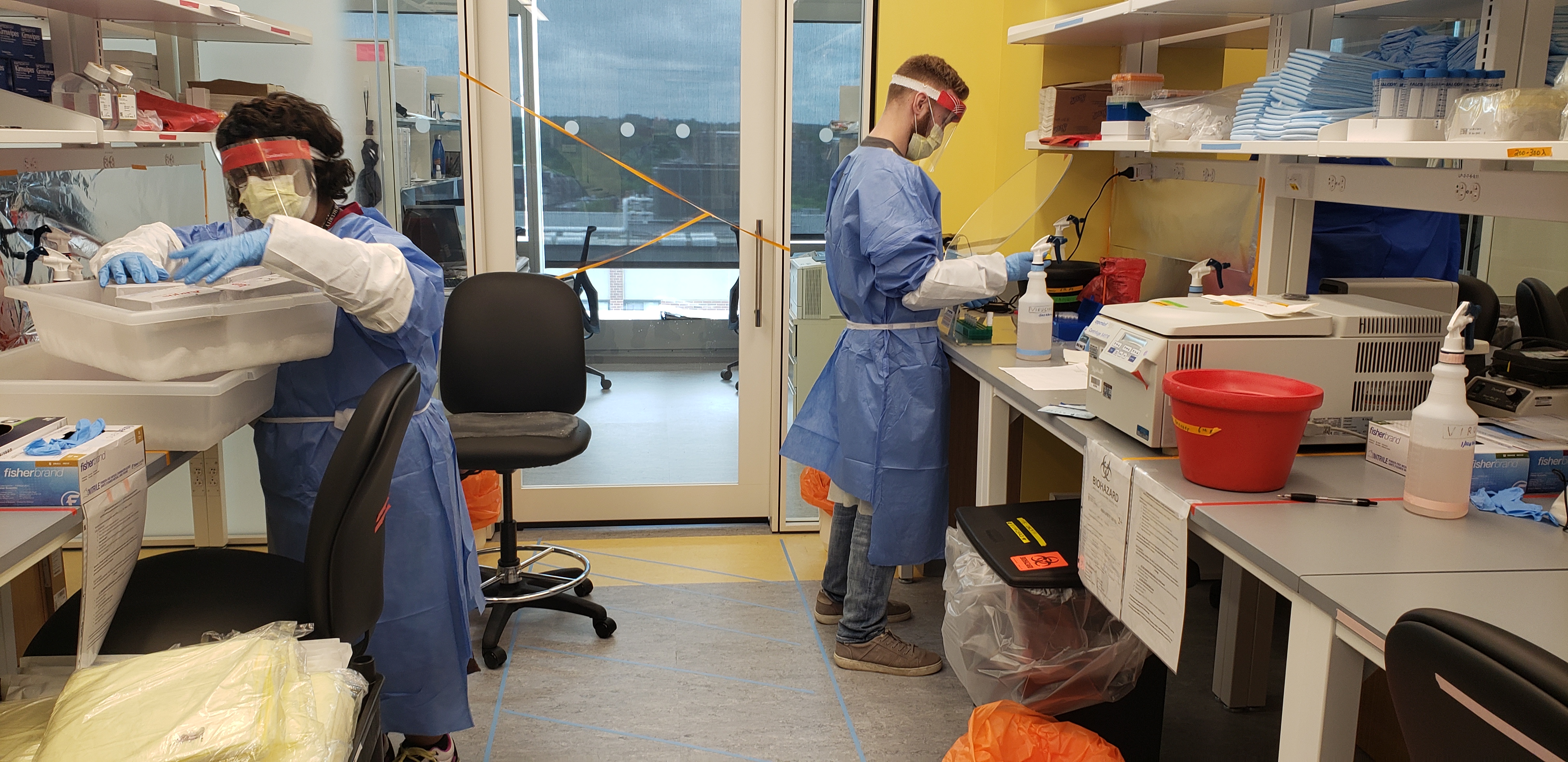
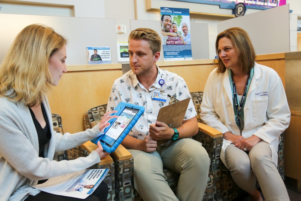
From left: Vannessa Davis and Samuel Bates support the MGB Center for COVID Innovation’s Diagnostic Accelerator.

With expertise that spans basic, translational and clinical research, Brigham clinicians and investigators have been working tirelessly to address the most urgent needs related to the COVID-19 pandemic.

To help reduce the spread of COVID-19, the Brigham shut down most of its physical research labs from March through the beginning of June. But even during the shutdown, many labs continued their work remotely and new efforts began to better understand the virus that causes COVID-19 (SARS-CoV-2), and the nature of the pandemic itself, in order to develop treatment strategies against them.

“In mid-March, hundreds of Brigham investigators and laboratory staff quickly pivoted to contribute to COVID-19 research and addressing COVID-related problems,” said Jacqueline Slavik, PhD, MSc, executive director of the Brigham Research Institute (BRI). “Within days, Brigham investigators were launching clinical trials, developing safer testing procedures and solving problems around personal protective equipment (PPE) shortages.”

Throughout the pandemic, Brigham investigators continued to pursue and receive funding for COVID-19 research — including clinical studies for a variety of potential therapies — through government and industry grants.

“Thanks to incredible ingenuity, a strong foundation and a breadth of core resources, our research community remains remarkably successful at putting forward award-winning research proposals and conducting first-class research,” said Paul Anderson, MD, PhD, chief academic officer and senior vice president of Research and Education. “As our research enterprise ramps up, these awards will become more important than ever, not only for individual research labs but also for the larger world as we continue to combat this pandemic.”

A Wave of New Solutions for COVID-19

Anesthesiologist Greg Crosby wears a 3D-printed face shield. Photo credit: Jim Rathmell, MD

Clinicians and researchers from the Brigham have come together with colleagues from around the world to produce a wave of innovative solutions faster than ever before.

Some of the Brigham’s advances in the fight against COVID-19 include:

  • Developing an innovative testing strategy to conserve PPE: A Brigham team developed the Brigham Protective Equipment for Clinical Test Environment and Diagnostics (B-PROTECTED) booth to preserve PPE and protect clinicians from COVID-19.
  • Creating in-house COVID-19 testing with results available within 24 hours: Brigham investigators implemented a rapid in-house test for COVID-19 patients who’ve been admitted to the Brigham but don’t yet have a definitive diagnosis.
  • Designing new face shields to protect health care workers from infection: A team of clinicians at the Brigham worked with academic and industry partners to design and develop a new 3D-printed face shield that offers a number of advantages over traditional shields.
  • Developing new protective materials: The lab of Jeff Karp, PhD,is working on an extended-duration sanitizer and a nasal spray to form a shield that protects against inhaled pathogens and viruses.
  • Investigating a safer way to split ventilators: Pulmonary physicians and biomedical engineers have been working together to develop a system that can be built from off-the-shelf components to allow for patient-specific volume and pressure control when using a single ventilator for more than one patient.
  • Using sewage to map an outbreak: Physician-investigators Peter Chai, MD, and Tim Erickson, MD, both of the Division of Medical Toxicology, are working with collaborators to develop technology and a plan for sampling sewage in North Carolina and Boston. These samples may provide important clues about the presence or absence of SARS-CoV-2 over time.
  • Investigating connections during social distancing: The lab of Amar Dhand, MD, DPhil, of the Department of Neurology, is researching the connections between individuals and their social networks. The team is learning firsthand how to stay connected during the time of social distancing.
  • Developing a universal coronavirus vaccine: The lab of Thomas Kupper, MD, chair of the Department of Dermatology, is investigating a vaccine that may protect against COVID-19 along with past, current and future strains of coronavirus.
Shriya Srinivasan and colleagues are working on a safer way to split ventilators

Shriya Srinivasan and colleagues are working on a safer way to split ventilators

“It’s inspiring to see how highly collaborative the Brigham research community has been during this crisis,” said Slavik. “Our research efforts have involved countless individuals from many academic institutions, the technology sector, industry and private companies — all of whom are working towards the common goal of mitigating COVID-19.”

Clinical Studies to Understand, Treat and Prevent COVID-19

To better understand COVID-19 and the virus that causes it, investigators are working on a range of studies and trials. These include:

  • Remdesivir clinical trials: The Brigham is a clinical trial site for evaluating the antiviral medication remdesivir in patients with COVID-19.
  • Learning from patients who’ve recovered from COVID-19: The lab of Duane Wesemann, MD, PhD, of the Division of Immunology and Allergy, is testing blood samples from people who’ve recovered from infection. These samples will help the team learn more about rates of exposure, the types of antibodies an infection elicits and the degree of immunity recovered patients have against re-infection.

“The Brigham has also created a COVID-19 biorepository to collect an array of biospecimens from patients who are or have been COVID-positive,” said Allison Moriarty, MPH, vice president of Research Administration and Compliance. “We believe this biorepository will be a key tool in helping us learn how to detect, treat and prevent COVID-19 in the future.”

A New Center for COVID Innovation

To rapidly develop new innovations and protect frontline staff across the Mass General Brigham (MGB) community and beyond, colleagues at Massachusetts General Hospital (MGH) and the Brigham research community launched the Mass General Brigham Center for COVID Innovation (MGBCCI) in March.

We create breakthroughs. It's in our DNA logo.“The mission of the MGB Center for COVID Innovation is to organize and consolidate the rapid investigation and clinical deployment of devices, diagnostics, data, analytics and the therapeutics that MGH and the Brigham is generating to combat the COVID-19 crisis,” center co-director said David Walt, PhD, a medical diagnostics researcher at the Brigham and Harvard University’s Wyss Institute for Biologically Inspired Engineering.

Investigators at the MGBCCI directly responded to, and continue to address, the most pressing needs that face health care workers — prototyping and testing new PPE, patient isolation hoods, alternative versions of respirators, face masks, face shields and nasal swabs.

Working groups within the MGBCCI have already identified and developed several new devices. They’re exploring other potential solutions for problems related to the pandemic. These efforts include:

  • Improving surgical mask design: The surgical masks group identified design inputs and criteria that are being applied to the design of an “ideal” surgical mask to improve upon the current design.
  • Reusing ventilators: The ventilators team is investigating the potential disinfection and reuse of HME/HEPA ventilator filters. They’re also working with software engineers to build a remote monitoring and alarm system for ventilators.
  • Replacing N95 respirators with novel devices: The N95 respirators team is exploring novel ideas for devices that could replace N95 respirators that aren’t dependent on the filtration media supply chain. They also developed a way to repair 50,000 defective N95 respirators with New Balance.

“We’re also working to identify a direct-to-consumer diagnostic test that could be used at home. When implemented, people can quarantine themselves if they are positive for COVID-19,” said Walt. “These tests could help stem flare-ups of COVID-19 cases that will invariably happen when people return to work and reintegrate into society.”

Leave a comment
Nancy Donovan

Nancy Donovan

The death of a spouse often means the loss of intimacy, companionship and everyday support for older adults. A new Brigham-led study finds that widowhood can have another profound effect: It may accelerate cognitive decline.

Investigators from the Brigham and Massachusetts General Hospital analyzed older, cognitively normal Americans enrolled in the Harvard Aging Brain Study whose marital status and brain β-amyloid levels — a marker of Alzheimer’s disease — were determined at the beginning of the study. The team found that individuals who were widowed experienced a sharper cognitive decline than their married counterparts, especially among those who had high β-amyloid levels.

The study suggests that widowhood may be an important and understudied risk factor for cognitive decline associated with Alzheimer’s disease and highlights the need for increased focus on this high-risk population. The team’s findings were published recently in JAMA Network Open.

“We know that social relationships can be an important buffer against cognitive decline,” said senior author Nancy Donovan, MD, of the Division of Geriatric Psychiatry. “Being married provides opportunity for more social engagement and emotional support from a spouse, it expands one’s social network and it provides more opportunity for cognitive stimulation. All of these benefits are lost in widowhood. Importantly, loss of a spouse is a highly stressful life event, which can have deleterious effects on the brain.”

Women are at increased risk for both widowhood and Alzheimer’s disease, both of which increase in frequency with age. The study, which included 260 cognitively unimpaired men and women ages 62 to 89, classified its subjects into three groups: married, widowed or unmarried (divorced, single, separated or never married).

We create breakthroughs. It's in our DNA logo.

Donovan and colleagues evaluated a participant’s cognitive performance each year for four years using a series of tests to analyze various dimensions of cognition. They found that cognitive performance declined in the widowed group, differing significantly from the married group. There was no difference between the married group and the unmarried group.

In addition, they found that among adults with the highest β-amyloid levels, widows had the sharpest decline in cognition compared to those who were married, declining at a rate three times faster. This finding was independent of many factors, including age, sex, socioeconomic status and a diagnosis of depression.

The authors noted this is the first study to show widowhood and β-amyloid have a combined effect on cognitive decline and will need to be replicated in other studies.
“Our division has become very interested in understanding the physiological effects of widowhood so that we can develop interventions to try to alter these trajectories,” said Donovan. “Our findings also suggest that researchers engaged in Alzheimer’s disease prevention trials may want to pay particular attention to widowed older adults to tailor interventions for this especially susceptible group of patients.”

1 Comment
Arash Mostaghimi

Arash Mostaghimi

In the largest study to date of skin cancer rates among gay, lesbian or bisexual individuals, a team of Brigham investigators found important differences in skin cancer prevalence among sexual minorities in the United States. Rates of self-reported skin cancer were higher among gay and bisexual men compared to heterosexual men, but lower among bisexual women than heterosexual women. The team’s results were published in JAMA Dermatology.

Arash Mostaghimi, MD, MPA, MPH, director of the Brigham’s Dermatology Inpatient Service, and colleagues analyzed data from the U.S. Centers for Disease Control and Prevention’s annual Behavioral Risk Factor Surveillance System (BRFSS) surveys from 2014 to 2018. The BRFSS interviews approximately 450,000 adults for the survey each year, and only began asking about sexual orientation and gender identity in 2014.

“This is the first time we’ve been able to look nationally at data about skin cancer rates among sexual minorities,” said Mostaghimi.

After analyzing data from 37 states, Mostaghimi and colleagues found that 8.1 percent of gay men and 8.4 percent of bisexual men had a history of skin cancer, compared to 6.7 percent of heterosexual men — a statistically significant difference. Women fared somewhat differently. Lesbian and heterosexual women had a similar rate of skin cancer (5.9 percent and 6.6 percent, respectively), while bisexual women had a considerably lower risk at 4.7 percent.

The survey did not collect information about risk factors for skin cancer. However, smaller studies have reported higher use of indoor tanning beds, a known risk factor for skin cancer, among gay and bisexual men.

We create breakthroughs. It's in our DNA logo.

The study has important implications for patient education and community-outreach initiatives for reducing skin cancer risk.

“This helps inform the nation about how to allocate health resources and how to train future physicians,” Mostaghimi said. “As a next step, we want to connect with sexual-minority communities to help identify the cause of these differences in skin cancer rates.”

The findings also reinforce the value of asking questions about sexual orientation and gender identity in national health surveys, he added.

“We have uncovered a clinically meaningful health variation,” Mostaghimi said. “If the BRFSS survey had never asked questions about sexual orientation and gender identity, we would never have known that these differences exist.”

Leave a comment
Michael Fischer

Michael Fischer

A new nationwide study led by investigators at the Brigham found that more than a quarter of antibiotic prescriptions dispensed to Medicaid recipients were not associated with a clinician visit. The results, published in Health Affairs, raise questions about the effectiveness of efforts to curb inappropriate prescribing.

A wide range of clinical guidelines aim to reduce the prescribing of antibiotics for illnesses not caused by a bacterial infection. These efforts aim to help lower the rate of antibiotic prescribing in the U.S., which is about double that of many other countries. However, such efforts may miss antibiotics that providers prescribe outside of clinical visits.

After evaluating nearly 300 million antibiotic prescriptions filled by Medicaid recipients over a decade, investigators found 28 percent were not associated with a clinician visit in the preceding seven days. In addition, 17 percent were prescribed after visits where no infection-related diagnosis had been documented.

“If we’re thinking about how to improve antibiotic prescribing, we need to understand the context in which it occurs,” said the study’s lead author, Michael Fischer, MD, MS, of the Division of Pharmacoepidemiology and Pharmacoeconomics. “If prescribing is taking place outside of an office visit, most of the approaches we are taking to combat antibiotics overuse will miss those completely.”

To capture these blind spots, Fischer and colleagues examined national Medicaid claims data from 2004 to 2013. They identified 298 million antibiotic prescriptions filled during this time for 53 million patients. The researchers used this data to determine whether patients had made a clinic visit in the seven days prior to filling the prescription. When there was a visit, they checked whether billing claims data specified an infection as the cause for the visit.

We create breakthroughs. It's in our DNA logo.

The authors acknowledge that a critical question remains: What occurred clinically in the many cases when antibiotic prescriptions were dispensed without a visit?

Since researchers’ analyses were based on claims data, they did not have access to medical records to determine what types of interactions took place between patients and prescribing clinicians. The authors said they assume most of these prescriptions were associated with a phone interaction, although some communication may have occurred over email, via web portals or during informal, uncaptured visits. Most encounters would be blind spots for existing interventions designed to improve appropriate antibiotic use, they noted.

Fischer said it will be important for future research to use clinical data to examine outcomes for patients who are prescribed antibiotics outside of office visits, adding that future interventions should be designed with the study’s findings in mind so that this large proportion of prescriptions is not missed.

Leave a comment
Juan Herrera-Escobar

Juan Herrera-Escobar

A new study led by investigators at the Brigham found that survivors of firearm violence have worse long-term outcomes than those who had sustained similar injuries in motor vehicle crashes. The results, published in Annals of Surgery, highlight the need to follow survivors after discharge and provide continued care.

The research team, led by investigators from the Brigham’s Center for Surgery and Public Health (CSPH), found that six to 12 months following a traumatic injury, rates of chronic pain, post-traumatic stress disorder (PTSD) and other poor physical and mental health outcomes were alarmingly high among survivors of firearm violence.

“When it comes to the public health problem posed by firearm injury, death is just one piece of the puzzle,” said corresponding author Juan Herrera-Escobar, MD, MPH, research director of Long-Term Outcomes in Trauma at CSPH. “Mortality rates for trauma patients have been dropping significantly over the last 20 years, but this presents a new challenge: What will we do for patients whose lives we save but who continue to suffer from the repercussions of traumatic injury? Our study shows that injury, and especially firearm injury, casts a long shadow over the lives of those who survive.”

As part of the Functional Outcomes and Recovery after Trauma Emergencies (FORTE) project — a study out of the CSPH that collects data from patients to understand the long-term recovery experience after traumatic injury — Herrera-Escobar and colleagues surveyed 63 adults who survived traumatic injury after being treated at one of three level-I trauma centers in Boston between 2015 and 2018. FORTE is intended to measure long-term outcomes that are meaningful to patients and their families; metrics include functional status, health-related quality of life and treatment adherence.

We create breakthroughs. It's in our DNA logo.

The team analyzed outcomes for people who had survived firearm injury and compared this data to those who survived similar injuries sustained in motor vehicle crashes.

Among survivors of firearm injury, 68 percent reported daily pain, 53 percent screened positive for PTSD, 39 percent reported a new functional limitation in an activity of daily living (such as walking, cooking, eating or going to the bathroom) and 59 percent had not returned to work. Most individuals experienced more than one of these negative long-term outcomes.

Not only were these rates significantly higher than those among the general population, but survivors of firearm injury were also far more likely than the survivors of motor vehicle crashes to have daily pain or PTSD and significantly worse quality-of-life issues related to physical and mental health.

The authors note that their findings may or may not be generalizable to the rest of the U.S. given their analysis was conducted at three urban trauma centers in Boston. As with all survey-based studies, this research may also be limited by selection and recall bias, as well as a lack of baseline information about those surveyed.

Herrera-Escobar said he and his colleagues have just begun to scratch the surface and more questions remain.

“We need to better understand the causes of these outcomes so that we can find opportunities for intervention,” he said. “This work has profound implications for trauma systems and highlights the limitations of their ability to help patients receive the proper services for a successful recovery after a traumatic firearm-related injury.”

Home

1 Comment

Brigham investigators found that abnormalities in a gene called SYCP2 were associated with low sperm count.

In a discovery 20 years in the making, Brigham investigators have uncovered a genetic abnormality that may be responsible for some unexplained cases of male infertility.

At least one in five cases of infertility remain unexplained. While men’s biology contributes to about half of these cases, doctors are often unable to identify a specific cause of their infertility. Researchers estimate that genetics could explain up to 50 percent of these cases, but many of the genes involved in male infertility remain unknown.

Now, Brigham investigators have found a chromosome rearrangement — a type of genetic mutation that alters DNA structure — and loss-of-function variants affecting one of those genes. Changes affecting this gene, known as SYCP2, are associated with low sperm count. The team’s findings, which report the first cases implicating this gene in four men with infertility, were recently published in the American Journal of Human Genetics.

“We hope that our evidence will contribute to this gene being included in panels for diagnosis of male infertility,” said medical geneticist and corresponding author Cynthia Morton, PhD, of the Department of Obstetrics and Gynecology and the Department of Pathology. “Infertility is a big problem for young people, and 40 to 72 percent of men lack a diagnosis. This means that we have a lot of gene-finding to do. My lab has a longstanding interest in studying individuals who have a balanced chromosome rearrangement where two chromosome segments swap places. In this case, it led us to an important discovery.”

The work that would lead the team to SYCP2 began in 1999, when Morton launched the Developmental Genome Anatomy Project (DGAP) to understand the genetic basis of birth defects and underlying molecular basis of development.

We create breakthroughs. It's in our DNA logo.

It was through this initiative that Morton and former graduate student and first author Samantha Schilit, PhD, along with colleagues from Harvard Medical School and Wesleyan University, were referred a case involving a 28-year-old man with a two-year history of infertility and severely low sperm count. By analyzing his chromosomes, the team found a genetic abnormality that led to a 20-fold rise in SYCP2 activity.

Through a series of elegant experiments involving yeast and cellular models, the researchers went about analyzing the effects of this abnormality, finding that it led to defects in sperm cell division — resulting in problems with sperm production and severely low sperm count.

“Balanced chromosomal rearrangements in infertile men are rarely followed up beyond reporting a risk for recurrent miscarriage. This work shows that a chromosomal rearrangement may also disrupt or dysregulate genes important in fertility, and therefore should be considered,” said Schilit.

In addition, the researchers looked for other cases of SYCP2 contributing to male infertility. To do so, they collaborated with investigators at the University of Münster in Germany who had enrolled men with infertility in a separate study. The University of Münster team’s search revealed three men with mutations that disrupt the function of SYCP2. These disruptions in SYCP2 were far more frequent among men with infertility than in the general population.

Morton notes that while the discoveries about SYCP2 may help inform a diagnosis, researchers still have much to learn about what it means for potential infertility treatments.

“A diagnosis can be therapeutic in itself — even if there isn’t something that can be done to correct it. It ends the search for the underlying issue and opens the door for enrolling in clinical trials,” said Morton. “And I believe there is good reason to be optimistic; we now have better tools for discovery and can begin on the path toward therapy.”

Home

Leave a comment
Dr. Suzuki speaking into microphone

Joji Suzuki speaks at a Discover Brigham panel.

On Nov. 7, members of the community interested in learning about the innovative research happening at the Brigham were invited to Discover Brigham for a full day of live demonstrations, speakers and scientific sessions. Attendees had the opportunity to hear from a Brigham researcher on a yet-to-be-identified plant that may be an eventual universal blood substitute for transfusions; immerse themselves in the future of Artificial Intelligence technology in medicine; and even get a look inside the medical bay on the International Space Station. We create breakthroughs. It's in our DNA logo.The event, hosted by the Brigham Research Institute, ended with an announcement that the $100,000 BRIght Futures Prize was awarded to Natalie Artzi, PhD, for her project aimed to fight childhood brain cancer.

Read more about Discover Brigham in the latest edition of Brigham Clinical & Research News.

Home

Leave a comment

Raul Piña-Aguilar

Cynthia Morton

When a couple loses a pregnancy, and especially when the same couple loses multiple pregnancies, doctors are often at a loss to explain why. For about 40 to 60 percent of couples who experience recurrent miscarriages, the condition remains unexplained, even after costly testing.

Chromosomal abnormalities — rearrangements of large chunks of DNA — in the genomes of one or both individuals trying to conceive are thought to be one of the major genetic causes of recurrent miscarriages, which is defined as having two or more pregnancy losses. Routine chromosome analysis can currently detect these abnormalities in about one in 50 couples. A new study by investigators from the Brigham, Shandong University and The Chinese University of Hong Kong describes a special genetic-sequencing technique, known as low-pass genome sequencing (GS), that can reveal chromosomal abnormalities in more couples than traditional testing — increasing detection to one in nine couples. The findings are published in The American Journal of Human Genetics.

“Recurrent miscarriages carry an underappreciated psychological and financial burden for affected couples. It’s often difficult to know how to treat or counsel couples when the cause of their infertility remains unknown,” said co-author Cynthia Morton, PhD, director of Cytogenetics at the Brigham. “There are couples who have chromosomal rearrangements that can’t be seen by classical methods. The technique we’ve used here increases the number of couples who we can detect who are at risk for having a miscarriage.”

Building on a previous collaboration, researchers from the Brigham worked with colleagues at The Chinese University of Hong Kong and Shandong University to analyze samples from nearly 1,100 couples who had two or more pregnancy losses. Couples were enrolled from recurrent miscarriage clinics at Hong Kong and Shandong universities. Upon performing both traditional chromosome analysis and low-pass GS on the samples, the team detected 127 chromosomal abnormalities using low-pass GS, accounting for approximately 12 percent of the couples. By comparison, traditional chromosomal analysis detected 86 chromosomal abnormalities.

We create breakthroughs. It's in our DNA logo.

“This a perfect example of the fundamental role that genetics can serve to help patients with infertility,” said co-author Raul E. Piña-Aguilar, MD, a postdoctoral fellow in the Morton lab.

“We have found that additional couples with recurrent miscarriages have chromosomal rearrangements. Undoubtedly, these couples are the ones who will benefit most from personalized intervention.”

A technique known as pre-implantation genetic testing can help couples with genetic abnormalities conceive through in vitro fertilization. Such approaches are expensive and, until now, it’s been unclear which patients with recurrent miscarriages would benefit most from genetic testing of their embryos.

“Our results suggest that applying low-pass GS could help identify a larger subgroup of patients at increased risk of subsequent miscarriages who might take advantage of pre-implantation genetic testing,” Morton said.


Home

Leave a comment

Over the last decade, evidence has mounted that the measles vaccine protects in not one but two ways: Not only does it prevent the well-known acute illness that frequently sends children to the hospital, but it also appears to protect the body from other infections over the long term.

How does this work?

Reporting on Oct. 31 in Science, an international team of researchers led by investigators at the Brigham and Harvard Medical School show that the measles virus wipes out 11 percent to 73 percent of the different antibodies that protect against other viral and bacterial strains a person was previously immune to — anything from influenza to herpes virus to bacteria that cause pneumonia and skin infections.

The study is the first to measure the immune damage caused by the virus and underscores the value of preventing measles infection through vaccination, the authors said.

“The threat measles poses to people is much greater than we previously imagined,” said senior author Stephen Elledge, PhD, the Gregor Mendel Professor of Genetics and of Medicine in the Blavatnik Institute at HMS and the Brigham. “We now understand the mechanism is a prolonged danger due to erasure of the immune memory, demonstrating that the measles vaccine is of even greater benefit than we knew.”

We create breakthroughs. It's in our DNA logo.

Elledge and colleagues found that those who survive measles gradually regain their previous immunity to other viruses and bacteria as they get re-exposed to them. But because this process may take months to years, people remain vulnerable in the meantime to serious complications of those infections.

As a result, researchers say clinicians may want to consider strengthening the immunity of patients recovering from measles with a round of booster shots of all routine vaccines, such as hepatitis and polio.

Answers in the Blood

This new discovery was made possible thanks to VirScan, a tool Elledge, George Xu and Tomasz Kula, doctorate students in the Elledge Lab, developed in 2015. VirScan detects antiviral and antibacterial antibodies in the blood that result from current or past encounters with viruses and bacteria, giving an overall snapshot of the immune system.

For the study, Elledge’s group used VirScan to measure antibodies before and two months after infection in blood samples from unvaccinated children who’d contracted the disease during a 2013 measles outbreak in the Netherlands. The researchers also compared the measurements to uninfected children and adults.

When Kula examined an initial set of these samples, he found a striking drop in antibodies from other pathogens in the measles-infected children that “clearly suggested a direct effect on the immune system,” the authors said. This was strong support for the immune-amnesia hypothesis previously put forward in 2015 from epidemiological data by Michael Mina, MD, PhD, who helped initiate the new study while serving as a postdoctoral researcher in Elledge’s lab. Mina, first author of the recent Science paper, is now an assistant professor of epidemiology at the Harvard T.H. Chan School for Public Health.

Further tests revealed that a severe measles infection reduced people’s overall immunity more than a mild case of measles. This could be particularly problematic for certain categories of children and adults, the researchers said, especially those who are malnourished or less healthy before contracting the virus.

Inoculation with the MMR (measles, mumps, rubella) vaccine did not impair children’s overall immunity. This aligns with decades of research.

Ensuring widespread vaccination against measles would not only help prevent the 120,000 deaths that will be directly attributed to measles this year alone but could also avert potentially hundreds of thousands of additional deaths attributable to the lasting damage to the immune system, the authors said.

“This drives home the importance of understanding and preventing the long-term effects of measles, including stealth effects that have flown under the radar of doctors and parents,” Mina said. “If your child gets the measles and then gets pneumonia two years later, you wouldn’t necessarily tie the two together. The symptoms of measles itself may be only the tip of the iceberg.”

Home

Leave a comment

William Kaelin displays a model of the VHL protein, which is involved in the groundbreaking research for which he was honored.

Early Monday morning, William G. Kaelin Jr., MD, a senior physician-scientist at the Brigham, received one of the most thrilling calls a scientist can get. The call from Stockholm revealed that Kaelin had been awarded the 2019 Nobel Prize in Physiology or Medicine.

“Like most scientists, I did occasionally dream that maybe one day this would happen,” said Kaelin. “I try to ignore that most days, but when I was younger, my late wife and I would have fun together talking about what would that be like. I’m accepting this prize partly on behalf of my late wife, Carolyn Kaelin. I like to think she’s smiling down and nodding.”

Kaelin, who is the Sidney Farber Professor of Medicine at Dana-Farber and Harvard Medical School, and a Howard Hughes Medical Institute Investigator, joined the Brigham in 1991 and currently has a research staff appointment in the Division of Medical Oncology in the Department of Medicine.

Kaelin is being honored jointly with Sir Peter J. Ratcliffe, MD, of the University of Oxford, and Gregg L. Semenza, MD, PhD, of Johns Hopkins University for their discoveries of how cells sense and adapt to oxygen availability. Their contributions and insights have paved the way for new strategies to treat diseases such as heart disease, anemia and cancer.

“As part of the Dana-Farber/Brigham and Women’s Cancer Center, we have great pride in the scientific research, discovery and innovation that occur at the Dana-Farber Cancer Institute. That pride extends to taking those basic discoveries and translating them into new advances for patients to improve their lives on a daily basis,” said Betsy Nabel, MD, president of Brigham Health. “There are broad applications for this oxygen sensing mechanism across many fields — heart and vascular disease, pulmonary disease, kidney disease and many other organ systems. Bill’s advances extend to cancer and far beyond, and for that, we are enormously grateful.”

The Path to Discovery

In his laboratory based at Dana-Farber, Kaelin’s work focuses on how mutations in genes known as tumor suppressors can lead to cancer. When Kaelin first established his lab, he read about the identification of a mutation in a tumor suppressor gene that leads to von Hippel-Lindau (VHL) disease. Patients with this rare, hereditary syndrome are at heightened risk for developing tumors in the kidneys, adrenal glands or pancreas.

We create breakthroughs. It's in our DNA logo.

Kaelin’s lab found that the VHL protein prevents the onset of cancer and helps control responses to low oxygen levels by regulating another protein that can trigger or suppress the production of red blood cells and the formation of new blood vessels. Cancer cells with VHL mutations can take advantage of this mechanism to surround themselves with new blood vessels that can fuel their growth. This process is known as angiogenesis and has become a target for combating many diseases, including cancer.

Oxygen sensing is essential for many different functions, including fetal development, metabolism, immune response, and exercise. When oxygen sensing goes amiss, it can lead to many diseases, including anemia, cancer, stroke, infection, heart attacks and more. Kaelin’s contributions to the fundamental understanding of how cells sense and respond to oxygen have led to the development of new drugs that can manipulate oxygen-sensing machinery to treat various diseases. Kaelin said he is continuing to search for new, promising targets.

“My mentor David Livingston, MD, taught me to think, ‘The most important thing you’re ever going to discover lies ahead of you,’” said Kaelin. “I still try to adhere to David’s advice.”


Home | Nobel Prize

8 Comments

bright futures 2019 prize

Now in its eighth year, the BRIght Futures Prize competition invites the Brigham community and the public to determine which of three investigators will be awarded $100,000 to support a groundbreaking research project designed to translate scientific discovery into clinical therapies.

Sponsored annually by the Brigham Research Institute (BRI), the BRIght Futures Prize supports Brigham investigators as they work to answer provocative questions or solve vexing problems in medicine. The winner is selected through a public vote, and Brigham faculty, staff and trainees are strongly encouraged to participate and help shape the future of innovation.

We create breakthroughs. It's in our DNA logo.

The finalists’ novel projects are featured in this special edition of Brigham Bulletin:

This year’s BRIght Futures Prize winner will be announced at Discover Brigham on Thursday, Nov. 7. Cast your vote at brightfuturesprize.org.


Home

Leave a comment
Senior leaders and scientists from Bayer, Brigham Health, Mass General and Partners HealthCare celebrate the new lab’s launch.

Senior leaders and scientists from Bayer, Brigham Health, Mass General and Partners HealthCare celebrate the new lab’s launch.

Scientists have studied lung diseases for decades, yet so much about them is still a mystery — including how they develop, why only certain people are afflicted with them and which treatments are most likely to be effective for any given patient.

Chronic lung disease is a broad term for several diseases of the airways and other structures of the lung, including chronic obstructive pulmonary disease (COPD). About 65 million people suffer from COPD and 3 million die from it each year, making it the third leading cause of death worldwide.

“The reality is that we don’t know what the key inciting events of COPD are, we don’t know how to reverse them therapeutically, and we’re not quite sure what the right molecular targets are,” explained Edwin Silverman, MD, PhD, chief of the Channing Division of Network Medicine. “We have a very rudimentary understanding of COPD. It’s an incomplete puzzle.”

We create breakthroughs. It's in our DNA logo.To help solve these unanswered questions and accelerate the search for lifesaving treatments, Brigham and Women’s Hospital, Massachusetts General Hospital and global pharmaceutical company Bayer announced the launch of a joint lab to research new drug candidates to treat chronic lung diseases. It will host scientists from all three parties, and Bayer is investing more than $30 million to fund joint research projects over the next five years.

Scientists in the new joint lab will work side by side, combining Bayer’s capabilities in drug discovery and development with the complementary clinical expertise, understanding of disease mechanisms, data-analysis capabilities and insights from leading physician-scientists at the Brigham and Mass General. More than 20 people from the three organizations will work in combined teams in the lab, located in the Brigham’s Thorn Building. The rights to the research findings will be shared equally between the organizations.

“We strongly believe that this model will significantly accelerate the pace of discovery toward the goal of getting new therapies from the lab to patients safely and efficiently,” said Paul Anderson, MD, PhD, senior vice president and chief academic officer at the Brigham. “This collaboration provides the opportunity to integrate novel findings directly into the drug development pipeline, thus speeding up the time to move a new treatment into the clinic.”

Brigham Health President Betsy Nabel, MD, also underscored the importance of joint efforts like this, noting that academic medical centers can no longer rely solely on diminishing government grants for research funding.

“Programs like this are incredibly important to sustain our research mission,” Nabel said during an event celebrating the joint lab’s launch on Sept. 24.

Within the framework of the new collaboration, four leading experts will combine their expertise in the search for new treatment options for patients who suffer from chronic lung diseases. In addition to Silverman, the team consists of Bruce Levy, MD, chief of the Brigham’s Division of Pulmonary and Critical Care Medicine, Benjamin Medoff, MD, chief of Pulmonary and Critical Care at Mass General, and Markus Koch, PhD, head of Preclinical Research, Lung Diseases at Bayer.

“This collaboration is a terrific match of long-standing clinical and research excellence at Brigham Health and Mass General with Bayer’s drug development strength,” said Chris Coburn, chief innovation officer of Partners HealthCare. “This state-of-the-art partnership has emerged from a shared commitment to improving patients’ lives.”

Home

Leave a comment
Terrie Inder headshot

Terrie Inder

When it comes to keeping the newborn brain healthy, taking steps to mitigate risk before birth may be critical. In ongoing investigations, clinical researchers from the Brigham are exploring whether a mother’s pomegranate juice intake during pregnancy can have a protective effect.

Some newborns, such as those with intrauterine growth restriction (IUGR) — a condition in which an unborn baby doesn’t reach a normal weight in the womb — are at heightened risk of brain damage. Being able to intervene before birth to aid in protecting the newborn brain may prevent the often-devastating effects of brain injury. In a recent paper in PLOS ONE, the Brigham team presented its preliminary findings from a clinical trial of expectant mothers whose babies were diagnosed with IUGR. The exploratory study, supported in part by an unrestricted gift from POM Wonderful, shows promise, with evidence of better brain development and brain connectivity in infants born to mothers who consumed pomegranate juice daily. A second, larger clinical trial is currently underway at the Brigham to validate these findings.

In cases of IUGR, a baby in the womb is measuring small for its gestational age often because of issues with the placenta, which brings oxygen and nutrients to the growing fetus. The process of birth
itself can further decrease blood flow or oxygen to the baby, including to the baby’s brain. If this is very severe, it can result in a serious, potentially fatal condition called hypoxic-ischemic injury.

Polyphenols are part of a class of antioxidants found in many foods and beverages, including nuts, berries, red wine and teas. Pomegranate juice is a particularly rich source of these compounds. Polyphenols are known to cross the blood-brain barrier, and studies in preclinical models have demonstrated protective effects against neurodegenerative diseases. To date, no clinical studies had evaluated the potential effects of giving pregnant women pomegranate juice to safeguard the brains of at-risk newborns.

We create breakthroughs. It's in our DNA logo.“Our study provides preliminary evidence suggesting potential protective effects for newborns exposed to pomegranate juice while in utero,” said senior author Terrie Inder, MBCHB, chair of the Department of Pediatric Newborn Medicine. “These findings warrant continued investigation into the potential neuroprotective effects of polyphenols in at-risk newborns, such as those with hypoxic-ischemic injury.”

The current randomized, controlled, double-blinded study enrolled 78 mothers from Barnes-Jewish Hospital obstetric clinic in St. Louis with IUGR diagnosed at 24–43 weeks’ gestation. Women were randomized to receive 8 ounces of pomegranate juice daily or a taste/calorie-matched placebo that was polyphenol-free. Women drank the juice daily from enrollment until delivery. The team measured several aspects of brain development and injury, including infant brain structure, minute change and functional connectivity.

While the team did not observe differences in brain macrostructure, they did find regional differences in white matter microstructure and functional connectivity.

“These measures tell us about how the brain is developing functionally,” said Inder. “We saw no difference in brain growth and baby growth, but we did see improvement in cabling network and brain development measured by synchronous blood flow and visual development of the brain.”

The authors note that the findings warrant the need for a larger, rigorously designed clinical trial to allow continued investigation into the potential neuroprotective effects of polyphenols. Such a study is now underway at the Brigham.

“We plan to continue investigating these exciting findings,” said Inder. “While the preliminary evidence shows promise, additional study and replication is needed.”


Home | PikeNotes | Brigham Values

Leave a comment

From left: Biobank team members Theodora O’Leary, Shane Sturtevant and Elizabeth Karlson meet at the Phlebotomy Lab.

Kristine Trudeau enrolled in the Partners HealthCare Biobank knowing she wanted to help advance medicine and potentially save lives. She never thought that her participation would eventually save her life, too.

Trudeau, a Brigham patient who lives in West Springfield, was recovering from a double lung transplant when she made the decision to join the Biobank — a long-term research program designed to help researchers and clinicians at the Brigham, Massachusetts General Hospital and other Partners institutions understand how genes, lifestyle and other factors affect people’s health and contribute to disease.

We create breakthroughs. It's in our DNA logo.

This spring, the Biobank surpassed 100,000 participants, making it one of the largest biobanks in the country helping to accelerate clinical research that will allow physicians to better understand, treat and prevent diseases now and in the future. As of today, the program has about 106,000 participants and is on track to reach 110,000 this fall.

After submitting a blood sample to the program, Trudeau, a geriatric home care and hospice nurse, was contacted by the Biobank and learned that genetic testing had revealed something unusual in her sample: a genetic marker that increases risk for breast, ovarian and prostate cancer. She opted to have her research result clinically confirmed and discussed the implications of clinical genetic testing with Carrie Blout, MS, CGC, a senior genetic counselor in the Brigham’s Genomes2People Program, which conducts research on the return of genomic information.

Following another DNA sample, Trudeau discovered she tested positive for a harmful variant in the BRCA2 gene, meaning she was at a high risk of developing breast or ovarian cancer. Blout encouraged her to meet with an oncologist, who recommended that Trudeau immediately begin aggressive monitoring for breast cancer and offered her the option to consider preventive surgical options, including having her ovaries and fallopian tubes removed.

As a result of the enhanced surveillance, Trudeau was diagnosed with breast cancer shortly thereafter. Fortunately, the cancer was found early, and she underwent a double mastectomy and oral hormone suppressant for treatment. In addition, she elected to have her ovaries and fallopian tubes removed. She is now in remission and credits her participation in the Biobank with helping her to take control of her health.

“I would have never known that I was at risk if I hadn’t donated to the Biobank,” said Trudeau. “I’d had a mammogram less than a year before and I had no issues. This information prompted the breast surveillance, and because I knew I was BRCA2-positive, once the cancer was detected, I opted to have the double mastectomy to help reduce further risks. If I hadn’t known this, I may not have made that decision. Having this information not only helped me. Because this is genetic, I was also able to encourage my sister to get tested, and at some point my children will be tested so that they can take precautions as well.”

Some Brigham-based members of the Biobank team, from left: Theodora O'Leary, Shane Sturtevant, Sherry Chen and Elizabeth Karlson

Some Brigham-based members of the Biobank team, from left: Theodora O’Leary, Shane Sturtevant, Sherry Chen and Elizabeth Karlson

‘Revolutionizing Research’

The Biobank has roots at the Brigham. The hospital’s former OurGenes, OurHealth, OurCommunity research program launched in 2009, merging with the Partners Biorepository for Medical Discovery at Mass General in 2013. The joint effort was renamed the Partners Biobank.

Participants provide a small blood sample that is linked to their electronic health record data as well as a self-reported health survey and their family medical history. After DNA research testing is performed, the samples and data relating to them are stored in a repository that is available to investigators at all Partners institutions. To date, the Biobank has provided samples or data to more than 200 studies.

“The Biobank has truly revolutionized the way we do research at Partners, and the more participants we have, the more powerful the resource is,” said Elizabeth Karlson, MD, of the Brigham’s Division of Rheumatology, Immunology and Allergy and one of the original leaders of the Biobank. “In addition to the sample size, a key differentiator for our Biobank is the electronic portal that we developed to help researchers search for disease phenotypes and request the data and/or samples they need for their studies.”

Driving Discoveries

Biobank researchers have been able to detect potential health issues for participants before any clinical symptoms were present. When a finding occurs, research geneticists refer patients to the appropriate clinical services at the Brigham, Mass General or Dana-Farber Cancer Institute. In some cases, participants knew that they had a personal or family history of the medical problem but did not know they were at genetic risk. In other cases, the Biobank was able to identify that participants were at risk of developing health concerns they had no prior knowledge of based on their personal or family medical history.

Scott Weiss, MD, principal investigator at the Biobank and scientific director of Partners HealthCare Personalized Medicine, said that greater participation in the Biobank enables Partners to increase the scale and scope of research and provides researchers with access to data and information that would otherwise take them years to source.

“This is a significant milestone for Partners and the research community,” he said. “We are already seeing tremendous results from the Biobank, both for individual patients with known health concerns to larger studies helping us identify diseases like Alzheimer’s and cancer in patients who have yet to develop any symptoms.”

To learn more about the Biobank or enroll as a participant, visit biobank.partners.org.


Home

Leave a comment
Mallika Mendu

Mallika Mendu

On July 10, the White House announced the signing of an executive order to launch Advancing American Kidney Health, a new initiative to improve the lives of Americans suffering from kidney disease, expand options for American patients and reduce health care costs. The new initiative builds on groundwork laid over the last several decades by many in the nephrology community, including leaders at the Brigham.

In this Q&A, originally published in Brigham Clinical & Research News, Mallika Mendu, MD, medical director for Quality and Safety at the Brigham, Director of Quality for the Brigham Renal Division, associate medical director at Partners Population Health for Specialty Programs and practicing nephrologist at the Brigham, shares her reflections on the initiative and its implications for providers and patients. For the last four years, Mendu has served on the American Society of Nephrology’s (ASN) Quality Committee, which, in collaboration with other nephrology and patient advocacy organizations, offered guidance that helped shape the Health and Human Services proposal leading to the new initiative.

What was your reaction when you heard this executive order was being signed?

MM: I felt a great deal of optimism for our patients with kidney disease that we as a nation are taking a huge leap forward in advancing kidney disease therapies and improving the quality of care delivered. I also feel a tremendous amount of pride as a member of the American Society of Nephrology, Partners Population Health Team and Brigham Renal Division — all of which have been championing efforts to transform kidney care delivery to improve outcomes for patients. It’s incredibly exciting to be able to share this news with my patients in clinic: Our government and our nation are paying attention to the need for advances in kidney health, and we have real reforms on the horizon.

Why is a comprehensive kidney health strategy to improve care for patients with chronic kidney disease so important?

MM: In comparison to cardiovascular disease and oncologic disease, there have been very few significant advances in kidney disease therapies for the past five decades. We are relying on technology (namely, dialysis) that was developed decades ago to treat patients with kidney failure. The last major legislative effort to help patients with kidney disease was the Medicare End Stage Renal Disease (ESRD) benefit in 1972. We now have 37 million patients with kidney disease, close to 700,000 with ESRD and Medicare costs over $35 billion a year attributable to the disease. It’s worth noting that ESRD accounts for less than 1 percent of the population, but over 7 percent of the expenditure. Finally, and most importantly, patients with ESRD have high rates of mortality, frequent utilization of care such as emergency department visits and hospitalizations, increased rates of disability and poor quality of life.

The comprehensive kidney health strategy, or Advancing American Kidney Health, facilitates improved patient outcomes, reduced medical expenditure and improved quality of life for patients. It’s a huge win for patients and for society.

What aspects of the announcement excite you most?

MM: I am excited that, for the first time, policy is focused on stemming progression of chronic kidney disease, increasing home dialysis, increasing transplantation and supporting innovation of new therapies. This contrasts with the traditional model of in-center hemodialysis. Many patients have voiced that in-center hemodialysis is taxing physically and emotionally. It’s also expensive for society to pay for the treatment through Medicare.

Can you tell us more about your involvement in the ASN’s Quality and Policy Committees and the team that outlined the population health initiatives?

MM: I have been extremely fortunate to have been given the opportunity when I was a fellow to serve first on ASN’s Public Policy Committee for two years. More recently, I’ve served on the ASN Quality Committee (for the past four years). It has been an absolute privilege and highlight of my career to have a voice on how policy is shaped and implemented. The Quality Committee comprises well-respected nephrologists across the nation who are actively engaged in clinical practice and most of whom are engaged in research efforts related to quality of care delivery for kidney disease patients. I have had the opportunity through ASN to meet with congressional staff on multiple occasions to advocate for kidney health policy and quality issues. Lastly, ASN partners with other kidney health organizations, including patient advocacy groups. Working closely with incredible patient advocates who share their compelling stories with congressional members has been inspiring.

In what other ways has the Brigham been involved in laying the groundwork for the new strategy?

MM: The Brigham has always been at the forefront of kidney health therapies and research. Dr. Joseph Bonventre, chief of the Renal Division, has championed basic science, clinical and translational research to advance the field, and has been incredibly supportive of public policy efforts. Our division has helped shape the national strategy that has resulted in Advancing American Kidney Health

We create breakthroughs. It's in our DNA logo.Dr. Bonventre has greatly encouraged the efforts to enhance awareness of kidney disease and expand resources for improvement of care and finding new therapies and was able to impact the public agenda of the ASN particularly during his time on the ASN Council and as its president. More recently he has chaired the Initiative to create a Technology Roadmap to Innovative Alternatives to Renal Replacement Therapy, which is intended to increase interest, investment and innovation in renal replacement therapy. This effort has contributed to the creation of the Kidney Innovation Accelerator (Kidney X), which is designed to foster innovation in preventing and treating kidney disease,

In addition, we have developed and implemented innovative approaches to care delivery for patients with kidney disease. We developed an Acute Kidney Injury (AKI) Standardized Clinical Assessment and Management Plan that guides providers on appropriate timing of renal replacement initiation for patients with AKI. We also developed a novel ESRD Care Coordination Program that seeks to reduce utilization (ED visits, hospitalizations) and facilitate transplantations for patients with ESRD, very much in the vein of what has been proposed. Finally, we’ve worked on the development of a chronic kidney disease (CKD) registry in concert with other Partners institutions to capture CKD patients across the network, identify key care delivery opportunities and identify patients at highest risk for developing ESRD.

As a physician who treats patients with kidney disease, what does the national attention to kidney health mean to you personally?

MM: In the clinic, I have had the chance to let my patients know about this exciting news. Several mentioned to me that they watched the announcement live and have hope that care delivery improvements are on the horizon. It also means a lot for kidney disease patients to know that their condition is not being ignored, their challenges are being recognized and there is now support for making things better. As a nephrologist who has focused her career on care delivery innovation and improving quality and safety, it’s a huge moment that carries a tremendous amount of promise.


Home

2 Comments
Raymond Mak headshot

Raymond Mak

Katelyn Atkins headshot

Katelyn Atkins

As advances in the treatment of non-small cell lung cancer (NSCLC) extend patients’ lives, more of these patients are facing a different threat: adverse cardiovascular events, such as heart attacks and heart failure.

Brigham and Dana-Farber Cancer Institute investigators led a new, retrospective study that examined outcomes for patients after receiving treatment for locally advanced NSCLC, finding that the average radiation dose delivered to the heart was associated with an increased risk of major adverse cardiovascular events and death. Among patients who did not have preexisting coronary heart disease, risk of having a major cardiovascular event after treatment exceeded the rates of people considered at high risk of such events. The team’s findings are published in The Journal of the American College of Cardiology.

“This is alarming data — to think that one in 10 of the patients I’m treating for this type of cancer will go on to have a heart attack or other major cardiac event,” said senior author Raymond Mak, MD, a thoracic radiation oncologist at the Brigham and Dana-Farber. “These cardiac events are happening earlier and more often than previously thought. More patients are living long enough to experience this risk of cardiac toxicity. We need to start paying attention to this and working together with cardiologists to help these patients.”

We create breakthroughs. It's in our DNA logo.

In many cases, a dose of radiation to the heart is the only way to treat a patient with lung cancer. Lung cancer is the leading cause of cancer deaths worldwide, and half of lung cancer patients will require radiation as part of their care. Previous studies have reported that advances in care, such as lung cancer screening and treating the disease with targeted therapies and immunotherapies, have improved survival rates. The average survival time is now more than two years for patients with locally advanced NSCLC.

“When treating patients with lung cancer, it’s a balance of risks,” said lead author Katelyn Atkins, MD, PhD, a resident in the Harvard Radiation Oncology Residency Program. “But we need to start thinking about where there’s room for improvement in optimizing treatment for patients and room for improvement in terms of collaborating with primary care physicians and cardiologists.”

To conduct their study, Mak, Atkins and colleagues analyzed data and outcomes for nearly 750 NSCLC patients treated with thoracic radiotherapy at the Dana-Farber/Brigham and Women’s Cancer Center and Dana-Farber Cancer Institute/Brigham and Women’s Hospital at Milford Regional Medical Center. After treatment, about 10 percent experienced a major adverse cardiac event, including heart attack and heart failure. The team saw increasing risk of cardiac events with higher dosages of heart radiation exposure, especially among patients who did not have coronary heart disease beforehand.

Based on their findings, the authors recommend more stringent avoidance of high-dose cardiac radiotherapy and suggest considering a much lower dose limit than national guidelines currently recommend.

“When possible, we should be thinking about ways to minimize cardiac radiation dose,” said Mak. “Recognizing that we may not always be able to do that, we’re now collaborating with our cardiology colleagues to explore early interventions to help mitigate the effects of cardiac injury from radiation therapy.”


Home

Leave a comment
From left: Sarah Thompson, Catherine Masse and Maureen Tapper discuss their research during a poster presentation.

Not only did Sarah Thompson (left) and Maureen Tapper (right) win funding during the Pitch Fest, but they also were honored for their poster, “A Lung Transplant Class: Improving Patient Care through Peer-to-Peer Education.” Additional authors for this poster are Catherine Masse (center), BSN, RN, PCCN, and (not pictured) Keri Townsend, PharmD, Karen Tsveybel, MSN, RN, CCTC, and Margaret Higgins, MSN, RN.

Following a high-energy pitch session before a live audience in Bornstein Amphitheater, two teams of nurses were named the winners of the inaugural Karsh Nursing Scholars Day “Pitch Fest” and received funding for novel initiatives designed to enhance care and the patient experience.

We create breakthroughs. It's in our DNA logo.

During the May 30 event, five nursing teams proposed original ideas for innovative tools, services and technologies. The two winning initiatives, “ICU Diary” and “Just Hanging Around: Bedside Organization,” will each receive $2,500 in funding via the Lily Kravitz Nursing Studies Award.

The pitch session was the culmination of Karsh Nursing Scholars Day, which honored Brigham nurses’ contributions to research, innovation and patient care. The event was made possible thanks to the generosity and vision of Estrellita Karsh, a longtime friend of the Department of Nursing and the hospital. The half-day celebration featured a poster presentation, demonstrations displaying innovations in nursing practice and drop-in sessions with Brigham nurse-scientists.

Maddy Pearson, DNP, RN, NEA-BC, chief nursing officer and senior vice president of Clinical Services at Brigham Health, noted during the pitch session that the diversity and depth of research highlighted the creativity and dedication of Brigham nurses.

Nadia Raymond (right) discusses her study with Sandra Parent.

Nadia Raymond (right) discusses her study with Sandra Parent.

“We are finding new, innovative ways to improve care, and we’re leaving no stone unturned,” she said.

The ICU Diary project — presented by Caroline Galligan, BSN, RN, Sharon Levine, MSN, RN, CVRN, and Melanie Nedder, MSN, RN, CCRN-CMC, of the Cardiac Intensive Care Unit (CCU) on Shapiro 9 East — will expand on a study the CCU conducted several years ago and implement diaries throughout all of the Brigham’s intensive care units to help patients remember their time in the hospital.

“Critical illness recovery is an emotional process,” said Nedder. “The ICU diary is a tool that fills gaps in patients’ recall of the ICU experience.”

Nedder and Galligan intend to roll out a diary prototype program by May 2020. They’re also designing comprehensive education for the ICU nursing staff who will implement the diaries at the bedside.

The Bedside Organization project — presented by Maureen Tapper, MSN, RN, PCCN, and Sarah Thompson, MSN, RN, CCNS, CWON, of the Thoracic Intermediate and Surgical Care Unit on Braunwald Tower 11ABD — proposed new and improved bedside organizers to hold patients’ call bells, mobile devices and other personal items.

Tom Tarbox and Debra Pelletier listen to a presentation.

Tom Tarbox and Debra Pelletier listen to a presentation.

“Currently, bedside tables are overused and not always accessible. Patients might not be able to find their call bells to get medication or go to the bathroom,” said Thompson. “There’s a potential for safety issues.”

Thompson and Tapper’s prototype is a single-use organizer that can safely attach to hospital-bed guard rails and stow valuables. These organizers will be piloted on Tower 11ABD, and staff will evaluate their effectiveness.

“This will improve patient satisfaction, care and safety, as well as help with loss prevention,” said Thompson.

Other innovative proposals featured during the event included a user-friendly iPad app to help lung transplant patients transition back to life at home, a bed-warming supply kit to improve infant care in the Neonatal Intensive Care Unit and a device designed to prevent central line-associated bloodstream infections in cardiac surgery.


Home

Leave a comment


In honor of Clinical Trials Day on May 20, faculty and staff from across the Brigham participated in a national campaign paying tribute to scientific advances propelled by clinical trials and raising awareness about career paths in clinical research. As part of the event, Brigham dietitians, investigators, laboratory technicians, physicians, nurses and research coordinators shared team photos and selfies with signs articulating some of the important contributions of clinical researchers.

At a Glance: Brigham Center for Clinical Investigation, 2018

    • 131 Active investigators

    • 245 Active clinical trials

    • 12,569 Clinical trial participant visits

Home

Leave a comment
Vesela Kovacheva snuggles with her oldest daughter, Kalina, moments after her birth at the Brigham.

Vesela Kovacheva snuggles with her oldest daughter, Kalina, moments after her birth at the Brigham.

As an obstetric anesthesiologist and a mother of two daughters, Vesela Kovacheva, MD, PhD, of the Department of Anesthesiology, Perioperative and Pain Medicine, has a keen understanding of what many of her patients experience during childbirth.

Most mothers undergoing a C-section receive a spinal anesthetic prior to the procedure — allowing them to remain comfortable while still being able to see and hear their newborns right away. But in nearly 75 percent of patients, it has an undesirable side effect of causing the mother’s blood pressure to drop. To counteract this, patients also typically receive another type of medication, a vasopressor, to elevate blood pressure.

This push and pull between the two drugs is a delicate dance as anesthesiologists work constantly to keep a mother’s blood pressure at a normal, controlled level before, during and after a C-section. If the numbers move too far in one direction or the other, mothers can experience nausea, vomiting, light-headedness and, in rare cases, stroke — events that may also have consequences for their baby’s well-being.

To keep patients safe and healthy, anesthesiologists monitor patients’ vital signs throughout the procedure, manually calculating and adjusting the medication dosage on an infusion pump, as needed, on a minute-by-minute basis — among multiple other tasks during surgery.

“The delivery of their baby is a very special time for our patients. It’s something they’re going to remember for the rest of their lives,” Kovacheva said. “Yet it’s also a very busy time for us as clinicians. We’re doing all of these things simultaneously while taking care of an anxious, awake patient and preparing for major abdominal surgery. In a couple of minutes, the math becomes too complex for any human to do, and we may under- or overtreat the patient.”

There must be a better way to do this, Kovacheva recalled thinking while recovering from her own C-section following the birth of her younger daughter at the Brigham two years ago.

We create breakthroughs. It's in our DNA logo.

It wasn’t long before she got to work on identifying and developing a possible solution using machine learning, a type of artificial-intelligence technology in which computers can be trained to identify patterns and make predictions after analyzing massive amounts of data.

Now, in collaboration with colleagues across the Brigham and Partners HealthCare specializing in anesthesiology, data science and pharmacology, Kovacheva and her team are working to develop a software algorithm that could monitor, anticipate and, under physician supervision, react to changes in maternal blood pressure in real time.

“We envision this as a physician superpower — something that augments, not substitutes, the doctor’s expertise. There will always be a need for a physician to tell the machine what to do,” she said. “Our hope is that this software will delegate a tedious, error-prone task to the machine so that I, as an anesthesiologist, could hold my patient’s hand, talk to them and focus on other aspects of their care.”

Training the Machine

While clinical guidelines provide a starting point for vasopressor dosage based on the particular clinical situation, every patient is unique and requires a personalized approach, Kovacheva explained. Patients who share similar traits may react to the same medication dose in different ways, and arriving at the right level is sometimes a matter of trial and error, she added.

The technology Kovacheva and her colleagues are developing would analyze a patient’s real-time blood pressure and heart rate and compare that against a large database of similar scenarios. In turn, the software will recognize when the medication needs to be adjusted and alert an anesthesiologist about the patient’s status and recommended dosage change.

Vesela Kovacheva

Vesela Kovacheva

“The way machine learning works is that it utilizes previous patient records and ‘teaches’ itself what should be done, based on what we’ve done in the past,” she said. “The most challenging part is that we need a very large number of patient records to train the software. We currently have about 700 records, and we’ll be doing a lot of internal testing to make sure that it’s safe and effective before we test it in people.”

If successful, Kovacheva hopes the technology could be useful to providers in more remote areas, where anesthesiologists are often in short supply, as well as in other types of surgeries and procedures.

For now, she is eager to do whatever she can to ensure childbirth is a joyous time and goes smoothly for more patients, families and providers alike.

“I feel that I have an idea that will hopefully make this experience better for not only moms and babies but also for the people caring for them,” Kovacheva said.

Home

Leave a comment
Sam Patz

Sam Patz

The speed of the human brain is remarkable — in a fraction of a second, neurons are activated, triggering thoughts and reactions to stimuli like sound or light. The speed at which we can noninvasively follow brain function using an MRI has not been as impressive, but a team of Brigham investigators hopes to change that.

An existing MRI method to measure brain function is known as functional MRI (fMRI) and is based on detecting changes in blood oxygen levels. While fMRI has revolutionized the field of neuroscience by showing which specific areas of the brain “light up” during different brain tasks, the changes that fMRI is sensitive to can take up to six seconds to be visible in humans — practically an eon in brain time.

Brigham investigators, in collaboration with colleagues at King’s College London and INSERM-Paris, have discovered a fundamentally new way to measure brain function using a technology known as magnetic resonance elastography (MRE), an approach that creates maps of tissue stiffness using an MRI scanner. In a recent paper published in Science Advances, the team presented data from preclinical studies indicating that the technique can track brain function activity on a time scale of 100 milliseconds. Studies of the technique in human participants are now underway.

“What excites me most is that this is an entirely new method, and I’ve always been intrigued by new science,” said co-corresponding author and physicist Sam Patz, PhD, director of the Low Field and Pulmonary MRI laboratories in the Department of Radiology.

Following the Clues

This work, which started out as a hunch and is now being borne out by rigorous experiments, represents the culmination of a five-year collaboration of an international team dedicated to the pursuit of this new way of imaging brain function.

Among those with whom Patz worked closely was co-corresponding author Ralph Sinkus, PhD, a physicist and professor at King’s College London and INSERM-Paris. A pioneer in the field of MRE, Sinkus played a key role in helping to launch the MRE research program for preclinical testing in Patz’s Boston-based lab and in carrying out the latest research. Both scientists point to each other as an example of how a true collaborative relationship should function.

Although Patz was initially interested in applying MRE to the lungs, the team decided to begin by scanning the brain. Their results revealed something surprising: The tissue in the brain’s auditory cortex, which processes sound, was stiffening for no apparent reason.

“These results were so unexpected that we had to pursue them, and this observation is what sparked everything else,” Sinkus said.

We create breakthroughs. It's in our DNA logo.On a hunch, Patz plugged one of the mouse’s ear canals with a gel. Sure enough, when he took another image, he saw the auditory cortex on the side of the brain that processed sound from that ear had begun to soften.

After this initial observation, the team spent more than a year modifying and improving both the MRE apparatus as well as the software to control the MRI scanner — both with the intent of improving the quality of the scans as well as designing a protocol to deal with issues they knew would be of concern to their colleagues. Then, with the new hardware and software, they rigorously demonstrated in preclinical studies that regions of the brain known to be activated by the stimulus they chose did, in fact, change their stiffness upon applying the stimulus. Researchers agreed the changes allow one to “see the brain thinking” in almost real-time.

“The intriguing novelty of this approach is that the stiffening/softening of specific brain regions persists even when stimuli as short as 100 milliseconds are presented to the mouse,” said Patz.

Next Steps

The team is now interested in using MREs to observe similar activity in the human brain, which could have implications for diagnosing and understanding diseases in which neuron activity may be slowed, disrupted or rerouted, such as Alzheimer’s, dementia, multiple sclerosis or epilepsy.

The team’s approach uses novel hardware to induce vibrations in the brain — an essential part to measure brain stiffness via MRI. Patz likens the apparatus to holding an electric toothbrush against one’s head to create tiny mechanical waves that travel through the brain.

The researchers presented data showing that altering the stimulus influenced the location, phase and intensity of the elasticity changes seen in the brain, meaning that they can visualize regional responses in the brain as they unfold at high speed.

“We believe this will transform our ability to observe neuronal functional activity with implications for neurological pathologies,” said Patz.

Leave a comment

With only months to live, Peter Bucciarelli was in desperate need of a double-lung transplant. When he heard about a new trial at the Brigham that would possibly increase his chances of receiving donor organs sooner, the 56-year-old patient didn’t think twice about enrolling.

This summer, he celebrates two years post-transplant. Bucciarelli is one of 35 patients who are thriving after participating in the DONATE HCV Trial, which studied whether thoracic organs (heart or lung) from donors infected with hepatitis C could be safely transplanted to recipients who did not have the virus. A multidisciplinary team of experts found they could successfully transplant the organs and then block the virus from replicating.

In a paper published in The New England Journal of Medicine earlier this week, the DONATE HCV Trial team describes a four-week antiviral treatment regimen started within hours of organ transplantation that prevented hepatitis C virus (HCV) infection in all patients, expanding the pool of eligible heart and lung donor organs. This is the largest clinical trial to date for HCV thoracic organ transplantation.

“There was a 100 percent success rate in terms of HCV treatment and six-month graft survival,” said corresponding author Ann Woolley, MD, MPH, of the Division of Infectious Diseases. “Direct-acting antivirals have revolutionized the field of hepatitis C treatment and have also created an opportunity to transplant organs from hepatitis C-positive donors.”

While transplants from these donors have been performed before, Woolley said the best approach to doing this — including when to initiate antiviral treatment, how long to treat patients after transplant and outcomes for heart and lung transplants — has not previously been systematically studied.

The Right Decision

From left: Ann Woolley and Peter Bucciarelli

From left: Ann Woolley and Peter Bucciarelli

Bucciarelli, a former contractor, said when his care team spoke with him about the trial, he knew it was the right decision to enroll.

“I was only given two months to live at the time,” said Bucciarelli, who was diagnosed with emphysema in 2010. “Being a part of this trial expedited things for me and ultimately saved my life.”

Since receiving his lung transplant, Bucciarelli has remained close with his care team, including Woolley, and often makes the four-hour drive from his home to the Brigham for routine checkups. He said he feels great and is thrilled to get back to living his life.

“I was able to see my daughter get engaged, and my son made me a grandfather for the first time,” Bucciarelli said. “Those are the moments I live for.”

Encouraging Results

In their paper, researchers presented data on patients who had enrolled in the study by February 2018. Six months or more after transplant surgery, all had undetectable amounts of hepatitis C and functioning transplanted organs. The team has enrolled 69 participants to date.

Nearly all patients who received organs from HCV-positive donors had evidence of the virus immediately after transplantation. However, very early, preemptive treatment prevented it from establishing an infection. All recipients cleared the virus within about two weeks, and it remained undetectable thereafter.

“It was critically important to us to determine this treatment not only prevented transmission of hepatitis C but also didn’t worsen outcomes for our transplant patients,” said co-author Steve Singh, MD, former surgical director of the Heart Transplantation and Mechanical Circulatory Support in the Department of Cardiac Surgery.

We create breakthroughs. It's in our DNA logo.

Although organ transplants in the U.S. have increased over the last five years, an estimated 1,000 patients die annually waiting for a lung or heart transplant. Drug intoxication deaths have led to a rise in available organs for transplantation, but donor hepatitis C infection has been a leading reason that otherwise medically suitable organs are deemed ineligible for transplantation.

“HCV infection has been a long-standing reason to decline donation of suitable organs,” said co-author Lindsey Baden, MD, director of Clinical Research in Infectious Diseases. “Transmission does occur, but a short, four week course of antiviral therapy led to rapid HCV clearance. These data demonstrate how preemptive therapy can stop transmission, thus decreasing medication burden, drug interactions and cost.”

The team also analyzed safety outcomes, finding that there were no hepatitis C-attributable adverse events.

“This study provided a unique opportunity to explore the utilization of thoracic organs from hepatitis C positive donors for transplantation, which to date have been underutilized despite being relatively common in the current donor population,” said co-author Hilary Goldberg, MD, MPH, medical director of the Lung Transplant Program. “This may allow us to provide successful transplantation to many recipients who might otherwise never have access to it.”


More About the Study

  • Standard treatment for people with a chronic hepatitis C infection is about eight to 12 weeks.
  • Other studies have found that it is feasible to treat kidney and liver transplant patients early after transplantation, and this approach is now being applied to heart and lung transplant recipients.
  • In this latest study, Brigham investigators set out to treat a much larger cohort of patients with a shortened, four-week therapy course and collected data on long-term outcomes.
  • The authors noted the importance of a shorter duration of antiviral treatment leading to successful outcomes for patients.
1 Comment
Front: Meir Stampfer and Francine Grodstein at the study’s biorepository. Back: Caitlin Barrows removes samples from a nitrogen freezer. (Photo by Stu Rosner)

Front: Meir Stampfer and Francine Grodstein at the study’s biorepository. Back: Caitlin Barrows removes samples from a nitrogen freezer. (Photo by Stu Rosner)

The following is an excerpt from the article “Connecting the Dots” in the latest issue of Brigham Health magazine. View the complete issue at brighamhealthmag.org.

Why do some people sail through life with few major illnesses while others begin experiencing chronic medical conditions in their 20s or even earlier? Your health relies on a unique combination of genetics, biology and other factors, including your habits, education and even ZIP code — from the air quality and walkability of your neighborhood to your access to health care.

Although there is no magic formula for a long, healthy life, decades of research confirm certain behaviors improve your chances: not smoking, staying physically active and maintaining a healthy diet. While these recommendations are practically common knowledge now, 50 years ago, that was not the case. Indeed, much of the health advice we hear today stems from a single source: the Nurses’ Health Study.

Brigham researchers started the study in 1976 with one goal: follow a large group of women over time to determine if oral contraception pills cause long-term effects on women’s health. When selecting a population of women to follow, the researchers thought nurses would be good participants because of their knowledge about health and reputation as a helping profession. On the first questionnaire mailed to 171,000 nurses across the U.S., 71 percent responded. This is almost unheard of in population studies, where response rates typically range from 5 to 25 percent.

Over the past 43 years, the study has surpassed its founders’ expectations, becoming the largest and longest running investigation of women’s health worldwide. It now includes 280,000 women and men across three generations.

Helen Levitan, who served as a nurse in Europe during World War II, was one of nearly 122,000 women who replied to the first mailing. For the next four decades until her death at age 96, she faithfully completed the biennial survey, answering questions about her health, diet, physical activity, medications and more.

Her daughter Jean remembers when Levitan, at age 92, complained she had not received the questionnaire that year: “My mom said, ‘They probably think I’m dead, but I want to fill out the survey!’ She was proud of being part of such a significant study, so we got the missing survey, and I helped her submit it.”

Gathering Clues

Levitan was not alone in her dedication to the study. Of the nurses who have joined, 90 percent continue to fill out questionnaires. “In population studies like this, it’s a dream to find such motivated participants because the more data we have, the more health conditions we can study,” said Francine Grodstein, ScD, director of the Nurses’ Health Study and an epidemiologist at the Brigham.

In addition to answering questionnaires, nurses provide biospecimens such as blood samples, saliva and nail clippings, which are stored in the BWH/Harvard Cohorts Biorepository and used to study how biology, genetics and lifestyle interconnect with health.

“It was a visionary decision by the study’s founders to start collecting biospecimens years before having the technology to analyze them in a sophisticated way,” said Edwin Silverman, MD, PhD, chief of the Channing Division of Network Medicine, which employs roughly 100 epidemiologists, biostatisticians and other Nurses’ Health Study staff. “With current technologies, we can generate data we couldn’t have dreamed of before, giving us an unprecedented opportunity to understand the biological causes of disease and better prevent, diagnose and treat medical conditions affecting so many people.”

Today, more than 1,000 researchers worldwide access data from the Nurses’ Health Study to conduct investigations. Their findings have identified many risk factors for chronic diseases, as well as ways to increase survival rates once a disease is diagnosed. They have also influenced laws and public health policies and recommendations, including U.S. guidelines on diet and physical activity.We create breakthroughs. It's in our DNA logo.

Connecting Nutrition and Disease

One of the study’s landmark findings in the early 1990s showed that consuming trans fats — mostly found in partially hydrogenated oils in processed foods — increases the risk of heart disease and death. Laws slowly evolved in response, starting with revised nutrition labeling for consumers. By June 2018, the U.S. Food and Drug Administration banned trans fats from the American food supply.

This achievement with trans fats came about primarily because of the work of Walter Willett, MD, DrPH, principal investigator of the Nurses’ Health Study 2. Since 1980, Willett has sent the nurses a supplemental food frequency questionnaire, which has revealed links between nutrition and many conditions, from kidney stones to cancer.

“We estimate more than 100,000 heart attacks will be prevented each year as a result of our finding about trans  fats,” said Meir Stampfer, MD, DrPH, an epidemiologist and longtime principal investigator for the Nurses’ Health Study. “Those 100,000 people won’t realize they’ve avoided a heart attack, and we won’t know who they are. Prevention does not get much attention in the public eye, but it is just as important as treatment, if not more so.”

Leave a comment
Jeffrey Sparks

Jeffrey Sparks

Smoking is an important risk factor for developing the most common form of rheumatoid arthritis (RA) and other inflammatory diseases, but a critical question remains: Can those who quit smoking delay or prevent RA, or have they permanently altered their risk of the disease? Brigham investigators recently tapped into data from the Nurses’ Health Studies to find out.

Their discoveries, which appear in Arthritis Care & Research, demonstrate for the first time that changing behavior — in this case, sustained smoking cessation — can reduce risk of developing seropositive RA, the more severe form of the disease.

“Ours is the first study to show that a behavior change can reduce risk for seropositive RA. Risk isn’t just about genes and bad luck; there’s a modifiable environmental component to the onset of this disease and a chance for some people to reduce their risk or even prevent RA,” said corresponding author Jeffrey Sparks, MD, MMSc, of the Division of Rheumatology, Immunology and Allergy.

To conduct their study, Sparks and colleagues used data from the original Nurses’ Health Study, established in 1976, and the Nurses’ Health Study II, a second cohort established in 1989. Both groups include female registered nurses from across the U.S. who filled out health surveys every two years with questions that included information about smoking and health status.

Among more than 230,000 participants in this analysis, the researchers identified 1,528 who developed rheumatoid arthritis. The team was especially interested in the 969 “seropositive,” as opposed to “seronegative,” cases. Patients with seropositive RA have related autoantibodies — a type of antibody that directs the immune system to attack healthy cells — and these individuals generally have more severe disease manifestations, including joint deformities and disability.

Among those with seropositive RA, risk began to decrease about five years after women quit smoking and continued to decline the longer they stayed nonsmokers. Participants who quit for good reduced their risk of the disease by 37 percent after 30 years.

The team did not find any association between smoking and seronegative RA, adding further evidence to the theory that seronegative and seropositive RA may be two distinct diseases with different risk factors but a similar clinical presentation.
We create breakthroughs. It's in our DNA logo.

“One of the lessons here is that it takes sustained smoking cessation to reap the full benefit,” said Sparks. “Whereas for other diseases, such as cardiovascular disease, quitting smoking can provide a more immediate effect, here we’re seeing benefits decades later for those who quit smoking permanently.”

While the biological mechanisms that link smoking and the development of RA are unclear, Sparks and others think that smoking may affect a preclinical disease process that leads to the formation of RA-related autoantibodies and increases inflammation.

In addition to further studying the biology of rheumatoid arthritis, Sparks and colleagues want to extend their investigations to include men and perform interventional trials among active smokers to see if it is possible to prevent the formation or lower levels of RA-related autoantibodies and the progression of the disease among those at increased risk.

Leave a comment