
Researchers hope to unlock brain’s mysteries with new MAGNUS MRI scanner
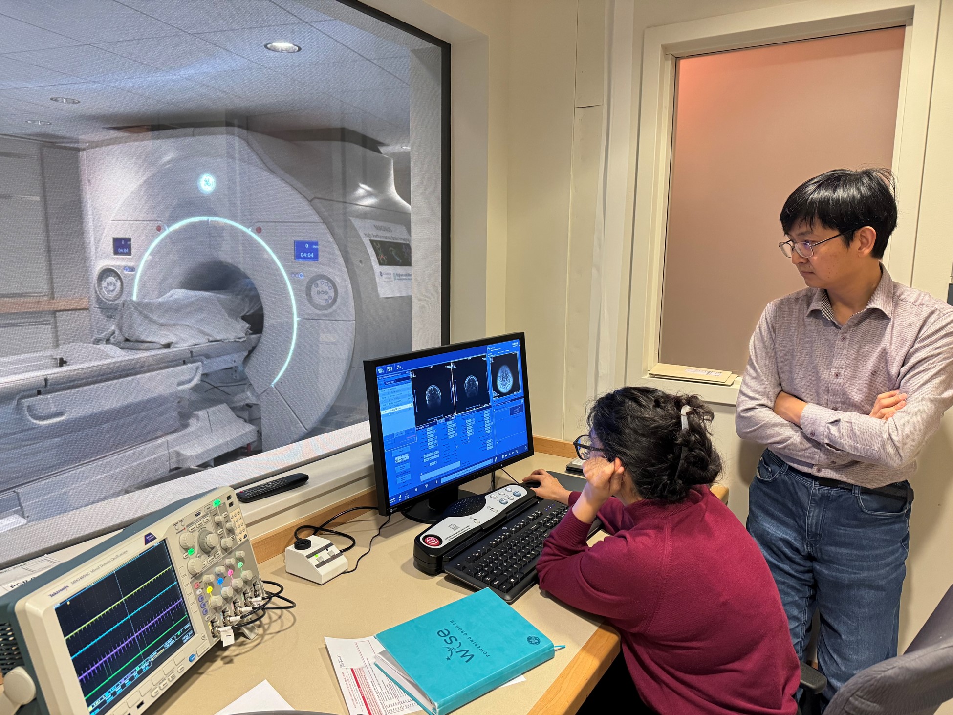
Nastaren Abad (left) and Seung-Kyun Lee (right) of GE HealthCare watch as the first human subject, a research volunteer, is scanned on the new MAGNUS MRI system at Brigham and Women’s Hospital.
One of the world’s most advanced neuroimaging devices — able to reveal the fine structure of the brain with an unprecedented level of detail — has been installed at Brigham and Women’s Hospital.
The Brigham is the fourth site in the world and the first in New England to acquire a MAGNUS scanner, an ultra-high-resolution MRI system that promises to reveal brain changes related to traumatic brain injury, Alzheimer’s disease, multiple sclerosis, brain tumors and many other neurological and psychiatric disorders. The Brigham system was delivered in late December and became operational in April. It was supported by a grant from the Massachusetts Life Sciences Center, with additional funding from Harvard Medical School and the hospital itself.
“With this system, we will be able to measure things that weren’t possible with conventional MRI,” said Carl-Fredrik Westin, PhD, the project’s principal investigator, who is also founding director of the Laboratory of Mathematics in Imaging and director of the Neuroimaging Analysis Center in the Department of Radiology. “We can ask questions we couldn’t ask before.”
MAGNUS is an acronym that stands for “Microstructure Anatomy Gradient for Neuroimaging with Ultrafast Scanning.” The head-only imaging system, which is installed in the MRI center at 221 Longwood Ave., is open to researchers across Mass General Brigham and the wider Boston area.
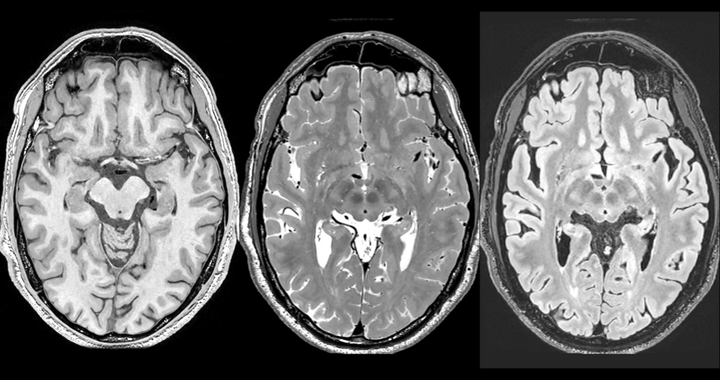
A research volunteer’s brain scan on the MAGNUS shows high-resolution images (0.7mm in all three axes) using three different protocols that are designed to reveal different aspects of brain structure.
While the MAGNUS is currently a research instrument, it nevertheless has great potential to advance future patient care.
“One of our near-term goals is to use it to study Alzheimer’s disease to try to find new biomarkers for the progression of disease,” explained Charles Jennings, PhD, executive director of the Program for Interdisciplinary Neuroscience and the Ann Romney Center for Neurologic Diseases. “Experts in the field believe Alzheimer’s starts to develop 10 or 20 years before patients are diagnosed, and we want to see those early changes to develop more effective treatments.”
According to radiology researchers, the MAGNUS will have a broad range of applications across neurology, neurosurgery and psychiatry. For instance, it can be used to study brain tumors, including the progression of gliomas, which researchers hope will improve clinical decision-making. Researchers also hope to visualize previously imperceptible differences in gray matter and white matter among people with schizophrenia and other psychiatric disorders.
“Also, because of its very high resolution, it can be used to map the white matter connections of the brain in ways that will be useful for surgical planning,” Westin said. “For example, when surgeons are planning to remove tumors or epileptic tissue, they try to avoid damaging healthy brain tissue. So, the more accurately you can map those boundaries, the better we can expect a patient’s quality of life to be after surgery.”

Leaders celebrate the arrival of MAGNUS with a ribbon-cutting ceremony at BWH on March 28. From left: Clare Tempany (vice chair for Radiology Research), Giles Boland (president of BWH and the Brigham and Women’s Physicians Organization), Paul Anderson (chief academic officer), Carla Reimold (Massachusetts Life Sciences Center), Caroline Shamu (associate dean, Harvard Medical School), Margaret Livingstone (HMS Neurobiology) and Carl-Fredrik Westin (BWH Radiology and PI for the MAGNUS project).
Another direction that researchers are excited to explore is measuring the flow of cerebrospinal fluid within the brain. This flow has been studied in animals, and it is believed to be important for clearing toxic waste products from brain tissue, but it was not possible to measure this in the living human brain using previous technologies. Researchers hope that the MAGNUS system, which is manufactured by GE HealthCare, will now make this possible.
“This is an exciting time for radiology research at the Brigham,” said Clare Tempany, MD, vice chair for Radiology Research. “We are looking forward to working with the GE team to explore the clinical potential of this powerful new instrument, and eventually bringing new treatments to our patients.”
Researchers interested in learning more about the MAGNUS system should contact Carl-Fredrik Westin, PhD, or Charles Jennings, PhD.